
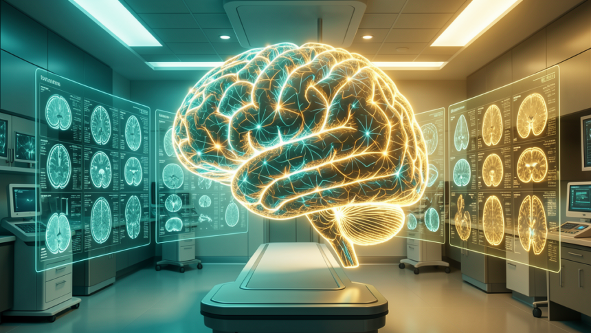
Researchers at Michigan Medicine have introduced NeuroVFM, a visual foundation model for neuroimaging that outperforms both frontier large language models and board-certified clinicians on brain scan interpretation, radiology report generation, and clinical triage. Published this week in Nature Medicine, the study represents a paradigm shift in how medical AI models are trained — and what they can achieve.
Trained on 5.24 Million Clinical Volumes
Unlike most medical AI systems that rely on curated research datasets, NeuroVFM was trained using what the researchers call a “health system learning” approach — drawing from 5.24 million clinical MRI and CT brain volumes spanning 566,915 studies collected over more than 20 years of routine clinical care at Michigan Medicine.
This uncurated, real-world training data includes the full spectrum of clinical presentations: common findings, rare pathologies, imaging artifacts, and the messy variability that characterizes actual hospital workflows. The result is a model that has learned from the same breadth of cases that experienced neuroradiologists encounter over decades of practice.
Beating Frontier LLMs at Their Own Game
In head-to-head benchmarks, NeuroVFM outperformed GPT-5 and Claude Sonnet 4.5 on neuroimaging interpretation tasks. More significantly, the model demonstrated substantially fewer hallucinated findings and critical errors compared to the frontier LLMs — a persistent problem that has limited the clinical deployment of general-purpose AI models in diagnostic radiology.
The performance gap highlights a fundamental insight: domain-specific models trained on authentic clinical data can surpass models trained on the entire internet. Where GPT-5 and Claude bring vast general knowledge, NeuroVFM brings deep, specialized understanding of how neurological pathology actually presents in clinical imaging.
From Narrow Tools to Generalist Diagnostic AI
Most FDA-cleared radiology AI products today are narrow, single-task tools — detecting one specific finding in one specific imaging modality. NeuroVFM points toward a different future: generalist diagnostic AI systems capable of interpreting complex imaging studies holistically, much as a human radiologist does.
The study also raises important questions about data access and institutional advantage. The “health system learning” paradigm requires access to massive, longitudinal clinical datasets that only large academic medical centers possess. How this approach can be democratized — or whether it will concentrate AI capabilities among a handful of elite institutions — remains an open question for the field.
For healthcare AI leaders, NeuroVFM reinforces a clear trajectory: the most impactful medical AI will be built not on internet-scale data but on clinical-scale data, trained within the walls of the health systems that generate it.