

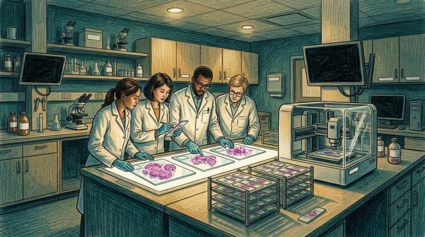
Cancer researchers at 10x Genomics will start reading tissue slides with help from Korean AI. The company is adopting Lunit SCOPE IO, an H&E pathology model, inside its oncology biomarker discovery work.
The model reads whole-slide H&E images. It characterizes tumor and stromal regions, immune-cell distribution and tertiary lymphoid structures in the tumor microenvironment. 10x will run that analysis next to spatial molecular data from its Xenium and Atera platforms.
The goal is to tie tissue morphology to the biology beneath it. 10x wants to surface biomarkers linked to treatment response and resistance.
The work will begin with clinical research in antibody-drug conjugates and immunotherapy response prediction.
“Lunit SCOPE IO adds the ability to analyze the full H&E landscape at scale,” said Brandon Suh, chief executive of Lunit.
Spatial methods reveal far more about a tumor and its surroundings, said Roman Yelensky, who leads clinical applications at 10x. He expects that molecular depth, combined with AI reading of the same routine slides pathologists use daily, to shorten the road to new biomarkers.
Lunit SCOPE IO is for research use only and is not cleared for diagnostic procedures. Lunit’s clinically validated portfolio is used at more than 10,000 sites across 65 countries, and its FDA-cleared breast screening suite sits in thousands of institutions. 10x Genomics, listed on Nasdaq as TXG, says its single-cell and spatial technologies appear in more than 10,000 research publications.
For cancer drug developers, the pairing targets a familiar bottleneck: linking what a pathologist sees on a slide to the molecular signals that predict who responds.