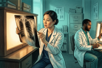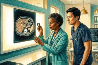


Millions of CT scans are ordered every day for chest pain, trauma or cancer staging. Bone density is rarely the reason. A new study in BMC Medical Imaging suggests that useful skeletal information is sitting unread inside those images, and that off-the-shelf AI can extract it.
Researchers at Changzheng Hospital of Naval Medical University in Shanghai tested whether a commercial deep learning tool could judge bone quality from routine non-contrast lumbar CT. They enrolled 518 patients who had undergone both a CT and the standard dual-energy X-ray absorptiometry, or DXA, scan, allowing a head-to-head comparison in the same people.
Each patient was sorted into one of three categories based on DXA: normal, osteopenia or osteoporosis. The AI, built on convolutional neural networks, then graded the CT images alone. The goal was not to replace DXA but to catch the many patients who never get one.
Why the screening gap matters
Osteoporosis is silent by design. Bone mineral drains for decades without pain until a hip, spine or wrist fracture suddenly changes a life. Effective drugs exist, but they only help once the condition is found. DXA is cheap, fast and safe, yet it is chronically underused, especially among older adults in primary care.
That is the case for opportunistic screening. If a scan already exists, asking software a second question costs almost nothing and adds no radiation and no appointment.
The Shanghai work is retrospective and single-country, so it is a proof of concept rather than a deployment. Still, it points to a practical shift: the value of routine imaging may lie as much in what it incidentally reveals as in the question it was ordered to answer.