

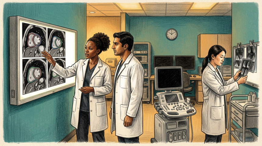
Ejection fraction decides a lot in cardiology: whether a patient gets a heart failure drug, an implanted device or surgery, and how closely they are watched. It is also notoriously reader-dependent.
A study in The Lancet Digital Health put an AI tool against experienced clinicians on 27,382 cardiac MRI scans from the UK and the United States. Patients were followed for more than five years on average, and 3,458 died. Measurements from the model predicted who was at higher risk of death slightly but significantly better than clinician measurements, and the gap survived adjustment for age and sex.
Seconds instead of thirteen minutes
The model took roughly two to three seconds per patient. Earlier work by the same team found expert manual analysis takes about 13 minutes. Errors were rare: a patient-level error rate near 1%, affecting 0.08% of images.
The work was led by City St George’s, University of London and University College London, with George Joy as lead author and Rhodri Davies, who invented the tool, as senior author. Funding came from an NIHR Clinical Lectureship, the NIHR UCLH Biomedical Research Centre and a British Heart Foundation accelerator award.
A first pass, not a verdict
The authors are explicit that clinicians still verify results and interpret scans in context. Modest accuracy gains compound across thousands of patients, but the error rate is a floor, not zero.
For stretched imaging services the practical claim is narrower than replacement: a fast, consistent first measurement, with expert review where the model is unsure. The team’s next step is combining MRI measurements with ECG data to catch muscle disease before damage sets in.