
Glioblastoma almost always comes back, usually near the cavity left by surgery. A team led by UC San Francisco and the University of Michigan has built a model that predicts where that first recurrence will appear, work published September 25 in Science Advances.
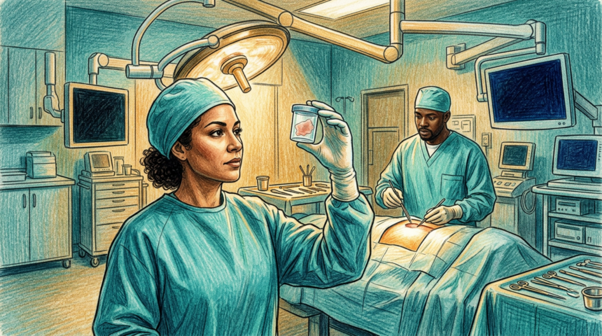
Conventional pathology needs dyes and stains, plus the handling time that comes with them. The team skipped that step. They imaged the surgical specimens with stimulated Raman histology, a technique that captures microscopic pictures of untouched tissue in under a minute, and ran those images through FastGlioma, a system built jointly at the two universities, to score tumor infiltration.
Training used roughly 300 samples from 60 patients; testing used about 100 samples from 20 more. Median time to recurrence in the cohort was 5.5 months. The infiltration score alone matched conventional pathology at flagging tissue that later gave rise to new tumor. Combined with clinical, imaging and molecular data across six machine-learning models, the best performer separated sites that did recur from those that did not, and infiltration was the strongest single predictor in five of the six.
The model localized recurrence well within 5 to 10 millimeters of sampled tissue.
What clinicians might do with it
If the signal holds, surgeons could remove more tissue during the initial operation where it is safe, or aim higher-dose focal radiation at predicted sites. Co-senior author Todd Hollon said the goal is delaying that first recurrence.