
A research team in India has taught an AI system to read colorectal cancer slides, and it borrowed optimization tricks from the animal kingdom to do it.
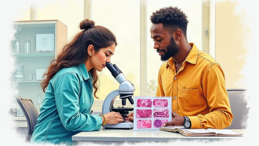
Histopathology is still the gold standard for diagnosing colorectal cancer, one of the world’s most common malignancies. Examining stained tissue under a microscope is slow and hard to scale. It is also vulnerable to differences between human readers, especially when tissue types look alike.
The new system, described in the journal Discover Artificial Intelligence, pairs two kinds of neural networks. A vision transformer called BEiT reads the whole slide first. It captures relationships between distant regions such as glandular structures, stroma, and cell nuclei. A convolutional network, EfficientNetB0, then refines the image for the final call.
The researchers found this cascade beat fusing the two models in parallel. The parallel design added roughly 60% more parameters without improving accuracy.
The bigger lift came from tuning. Rather than hand-pick settings, the team applied two nature-inspired algorithms. One mimics humpback whales spiraling around prey. The other models a flock that shares its best findings.
The result was a clear gain. Before tuning, the model scored 92.13%. After, it reached 96.27%.
The work was led by Hemanth K S, Samiksha Sandeep Zokande, and Prathana Sharma of CHRIST University in Bangalore, with N Kartik of the Manipal Academy of Higher Education.
The team frames the approach as a step toward cheaper, more consistent cancer screening. The model still needs validation across hospitals and patient populations before routine use.