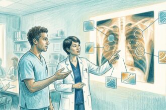

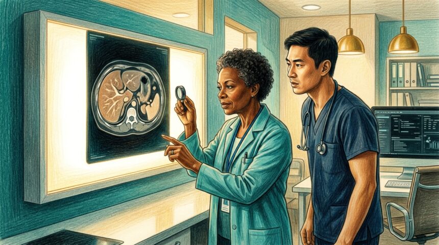
A new deep learning framework called PRISM can diagnose focal liver lesions on MRI scans taken without contrast dye, skipping gadolinium altogether.
Developed and validated on a multicenter cohort of 12,823 patients from nine institutions, PRISM works in three stages: automated lesion detection and segmentation, benign-malignant classification, and a four-subtype classification model for suspicious malignancies.
Across five test cohorts, the binary and four-subtype models achieved mean accuracies of 0.981 and 0.859. In a multireader study, 13 radiologists of varying experience gained accuracy with AI assistance, improving benign-malignant diagnosis by 4.4% and malignant subtype classification by 12.1%, while cutting interpretation time by 30.5% and 66.6% respectively.
A single-center prospective study of 1,147 consecutive patients showed PRISM’s triage value, identifying 79.3% of the cohort as low-risk with a 99.1% negative predictive value.
The work appears in npj Digital Medicine. Plain MRI diagnosis matters because contrast-enhanced scans are costly, time-intensive, and carry gadolinium burden, while ultrasound sensitivity is limited. Expert-level interpretable diagnosis on non-contrast MRI offers a viable path to streamline liver cancer workflows.