
Brain-computer interfaces have an imaging problem. Every patient’s speech and motor regions sit in slightly different places, so picking a neural target is guesswork dressed as surgery. Paradromics and Siemens Healthineers said on October 19 that they will build one imaging pathway to solve it.
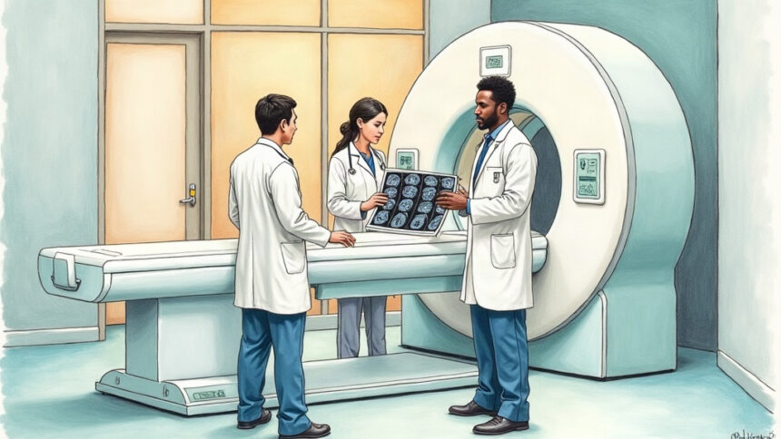
The collaboration joins Siemens’ 3T MRI hardware, including the MAGNETOM Cima.X, with Paradromics’ clinical work on its Connexus device, an implant designed to restore speech for people paralysed by injury or disease.
Seven steps, one workflow
The intended package runs the full arc: assessing whether a patient is a candidate, identifying the neural target, planning the surgical trajectory, navigating during the operation, confirming where the device landed, checking imaging predictions against real neural recordings, and tracking outcomes afterwards.
It leans on four modalities – structural MRI, functional MRI, vascular imaging and CT – so surgeons can see anatomy, find functionally active tissue and steer clear of vessels on the way in.
Matt Angle, Paradromics founder and chief executive, argued that hardware alone does not determine performance. Understanding each patient’s individual organisation, he said, is what lets a team place a device accurately.
Built for hospitals that already own scanners
William Marks, Paradromics’ chief clinical officer, made the commercial logic plain. Most US hospitals run Siemens systems, so the plan avoids asking them to buy and train on bespoke capital equipment.
Clinical investigators including Mass General Brigham’s Center for Neurotechnology and Neurorecovery will help shape the workflow. The first focus is speech and motor function, with hand control and post-stroke language restoration as possible extensions.
Connexus remains investigational and is limited by US law to research use. Paradromics recently reported four years of stable neural recording from its devices.